


- tel010-4724-3240
- fax02-447-3074
- time9시-18시

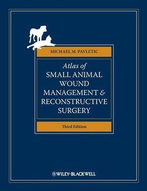


[P0000CRL] Atlas of Small Animal Wound Management and Reconstructive Surgery, 3rd Edition 


() 해외배송 가능
| 판매가 | |
|---|---|
| 소비자가 | 200,000원 |
| 적립금 |
|
| 무이자할부 | |
| 제조사 | |
| 원산지 | |
| 상품코드 | P0000CRL |
| 수량 |
|
| 국내/해외배송 | |
| SNS 상품홍보 | |
| QR코드 |
|
| QR코드 보내기 |
   
|


event
상품상세정보
VIN.com의 북리뷰에서 별 4개를 받았읍니다.
| Atlas of Small Animal Wound Management and Reconstructive Surgery, Third Edition Michael M. Pavletic, DVM, DACVS |
| Review by Kathy Lyon (Click on stars for an explanation) |
Reading this book, I can only wish you the good fortune to never have to see any of these cases, but we all know you will eventually. When you do, you will want this book.
This edition has added forty plates including sections on bandage/splint techniques, reconstructive surgery of the prepuce and the management of problematic skin fold conditions.
The book has also updated the section on management of problematic wounds and wound care products. Important points are boxed in the text for emphasis.
The book begins with a primer on the skin, starting with structure and continuing through circulation and congenital skin disorders. A map of surface and superficial arteries and veins is included. A valuable reference for repairs or grafts.
"The Basic Principles of Wound Healing" is a must-read before you attempt reconstructive surgery. The etiology of the wound is an important consideration in selecting the method of repair. Species variations are also to be considered. "Wound Management" follows, including presentations, assessing the wound, wound classification, selecting the proper closure technique and basic wound management in six simple steps.
There are important procedures to follow for selecting the proper closure technique. The various methods of closure and management are well illustrated and described.
There is a variety of injuries (vehicle damage, burns, gunshot wounds, dogfight injuries) illustrated, and a variety of wound care products listed as well as tips for their use. Topics of particular interest would include: non-healing wounds, exposed bone, inhalation injuries, porcupine quills, skin stretching techniques, use and creation of skin flaps, grafts, facial reconstruction, foot pad reconstruction, eyelid reconstruction, cosmetic closure techniques, and much more. I would suggest having a look at the table of contents on the Wiley-Blackwell site for encouragement. The topics are varied and detailed and will most likely convince you to buy the book. The accompanying photos are excellent and are directed toward helping the clinician through the surgery. The photos will reassure you that even those wounds that appear hopeless have promise.
The excellent photos are accompanied by instructive drawings. Surgical and repair techniques are addressed and illustrated.
Reading the book in advance of need is recommended. When that terrible-looking injury comes in the door, just knowing there is possibility of repair is a comfort to both the owner and the veterinarian.
Wiley-Blackwell (2010). 680 pages, hardcover. ISBN: 978-0-8138-1124-6.
Atlas of Small Animal Wound Management and Reconstructive Surgery, Third Edition is a full-color atlas that maintains the surgical focus of earlier editions while now presenting essential information on basic principles of wound healing, wound management, and common wound complications. The new edition presents a wider variety of topics including skin fold disorders, urogenital surgery, new flap techniques, and an expanded chapter on facial reconstruction. It also features 40 new plate illustrations, new sections on bandage and splint techniques, and significant updates on wound healing physiology, equipment, and dressing materials.
![]()
-
Full-color, one-stop reference that maintains the surgical focus of earlier editions while now also presenting essential information on basic principles of wound healing and wound management, common complications, and general surgical considerations
-
Discusses techniques for specific body areas such as the face, mouth, eye, nose, and foot
-
Presents a wide variety of topics including skin fold disorders, urogenital surgery, new flap techniques, and an expanded chapter on facial reconstruction
-
Contains 40 new plate illustrations, including new sections on bandage/splint techniques, reconstructive surgery of the prepuce, the management of problematic skin fold conditions, and features significant updates on wound healing physiology and equipment and dressing materials
-
Essential purchase for veterinary surgeons, residents, and small animal practitioners
![]()
1. The Skin.
Skin Function.
Skin Structure.
Cutaneous Adnexa.
The Hypodermis.
Cutaneous Circulation.
Congenital Skin Disorders.
2. Basic Principles of Wound Healing.
Introduction.
Wound Healing.
Species Variations in Wound Healing.
Artificial Skin.
3. Basic Principles of Wound Management.
Introduction.
Patient Presentation.
Mechanisms of Injury and Wound Terminology.
Wound classification.
Options for Wound Closure.
Healing by Adnexal Re-epithelialization.
“Pointers” in Selecting the Proper Closure Technique.
Basic Wound Management in Six Basic Steps.
4. Wound Care Products and Their Use.
Wound Drainage Systems.
Topical Wound Care Products.
Alternative Forms of Wound Therapy.
Concluding Remarks.
Plate 1: Vacuum-Assisted Closure.
5. Dressings, Bandages, External Support, and Protective Devices.
Introduction.
Composition of a Bandage.
Preventing Bandage Displacement.
Tie-over Dressing/Bandage Technique.
Pressure Points: Bandage Options.
Bandage “Access Windows”.
Bandaging Techniques for Skin Grafts.
Bandaging Techniques for Skin Flaps.
Splints, Casts, Reinforced Bandages
Miscellaneous Protective Devices.
Plate 2: Basic Bandage Application for Extremities.
Plate 3: Tape Stirrups and Padding Dos and Don’ts.
Plate 4: Elasticon Bandage Platforms and Saddles.
Plate 5: Spica Bandages/Splints.
Plate 6: Schroeder-Thomas Splint.
Plate 7: Schroeder-Thomas Splint: Security Band Application.
Plate 8: Body Brace.
6. Common Complications in Wound Healing.
Improper Nutritional Support.
Hypovolemia and Anemia.
The Nonhealing Wound: General Considerations.
Failure to Heal by Second Intention.
Scarring and Wound Contracture.
Infection.
Draining Tracts.
Use of Tourniquets for Lower Extremity Procedures.
Seromas.
Hematomas.
Exposed Bone.
Wound Dehiscence.
7. Management of Specific Wounds.
Bite Wounds.
Burns.
Inhalation Injuries.
Chemical Burns.
Electrical Injuries.
Radiation Injuries.
Frostbite.
Projectile Injuries.
Pressure Sores.
Hygroma.
Snakebite.
Brown Recluse Spider Bites.
Porcupine Quills.
Plate 9: Pipe Insulation Protective Device: Elbow.
8. Regional Considerations.
The Canine Profile.
The Feline Profile.
Plate 10: Surgical Technique Menu.
9. Tension Relieving Techniques.
Introduction.
Skin Tension in the Dog and Cat.
Plate 11: Tension Lines.
Plate 12: Effects of Skin Tension on Wound Closure.
Plate 13: Patient Positioning Techniques.
Plate 14: Undermining Skin.
Plate 15: Geometric Patterns to Facilitate Wound Closure.
Plate 16: V-Y Plasty.
Plate 17: Z-Plasty (Option I).
Plate 18: Z-Plasty (Option II).
Plate 19: Multiple Z-Plasties.
Plate 20: Relaxing/Release Incisions.
Plate 21: The “Hidden” Intradermal Release Incision.
Plate 22: Multiple Release Incisions for Extremity Wounds.
Plate 23: Walking Suture Techniques.
Plate 24: Skin Stretchers to Offset Incisional Tension.
Plate 25: “Tension” Suture Patterns.
Plate 26: Retention Sutures.
Plate 27: Stent.
Plate 28: Skin “Directing” for Maximum Coverage.
Plate 29: Relaxing Incision to Reduce Flap Tension.
10. Skin-Stretching Techniques.
Physiology of Skin Stretching.
Presuturing.
Skin Stretchers.
Skin Expanders.
Plate 30: Presuturing Technique.
Plate 31: Application of Skin Stretchers.
Plate 32: Skin-Stretcher Substitution for Presuturing.
Plate 33: Skin Expanders.
11. Local Flaps.
Introduction.
Advancement Flaps.
Rotating (Pivoting) Flaps.
Plate 34: Single Pedicle Advancement Flap.
Plate 35: Bipedicle Advancement Flap.
Plate 36: Transposition Flap (90 degrees).
Plate 37: Transposition Flap (45 degrees).
Plate 38: Interpolation Flap.
Plate 39: Rotation Flap.
Plate 40: Forelimb Fold Flap.
12. Distant Flap Techniques.
Distant Flaps.
Direct Flap.
Indirect Flap.
The Delay Phenomenon.
Plate 41: Direct Flaps: Single-Pedicle (Hinge) Flap.
Plate 42: Direct Flap: Bipedicle (Pouch) Flap.
Plate 43: Indirect Flaps: Delayed Tube Flaps.
13. Axial Pattern Skin Flaps.
Introduction: Axial Pattern Flaps.
Island Arterial Flaps.
Reverse Saphenous Conduit Flap.
Secondary Axial Pattern Flaps.
Plate 44: Four Major Axial Pattern Flaps of the Canine Trunk.
Plate 45: Skin Position and Axial Pattern Flap Development.
Plate 46: Omocervical Axial Pattern Flap.
Plate 47: Thoracodorsal Axial Pattern Flap.
Plate 48: Lateral Thoracic Axial Pattern Flap.
Plate 49: Superficial Brachial Axial Pattern Flap.
Plate 50: Caudal Superficial Epigastric Axial Pattern Flap.
Plate 51: Cranial Superficial Epigastric Axial Pattern Flap.
Plate 52: Deep Circumflex Iliac Axial Pattern Flap: Dorsal Branch.
Plate 53: Deep Circumflex Iliac Axial Pattern Flap: Ventral Branch.
Plate 54: Flank Fold Flap: Hind Limb.
Plate 55: Genicular Axial Pattern Flap.
Plate 56: Reverse Saphenous Conduit Flap.
Plate 57: Caudal Auricular Axial Pattern Flap.
Plate 58: Superficial Temporal Axial Pattern Flap.
Plate 59: Lateral Caudal (Tail) Axial Pattern Flap.
14. Free Grafts.
Free Skin Grafts.
Classification of Free Grafts.
Graft Thickness.
Partial Coverage Grafts.
Dermatomes.
Preservation by Refrigeration.
Intraoperative Considerations.
Bandaging Technique for Skin Grafts.
Plate 60: Punch Grafts.
Plate 61: Pinch Grafts.
Plate 62: Strip Grafts.
Plate 63: Stamp Grafts.
Plate 64: Sheet Grafts.
Plate 65: Dermatome: Split-Thickness Skin Graft Harvesting.
Plate 66: Mesh Grafts (With Expansion Units).
Plate 67: Mesh Grafts (With Scalpel Blades).
15. Facial Reconstruction.
Introduction: Facial Reconstructive Surgery.
Plate 68: Repair Of Lower Labial Avulsion.
Plate 69: Repair of Upper Lip Avulsion.
Plate 70: Wedge Resection Technique.
Plate 71: Rectangular Resection Technique.
Plate 72: Full-Thickness Labial Advancement Technique (Upper Lip).
Plate 73: Full-Thickness Labial Advancement Technique (Lower Lip).
Plate 74: Buccal Rotation Technique.
Plate 75: Lower Labial Lift-Up Technique.
Plate 76: Upper Labial Pull-Down Technique.
Plate 77: Labial/Buccal Reconstruction With Inverse Tubed Skin Flap.
Plate 78: Skin Flap for Upper Labial And Buccal Replacement (Facial Axial Pattern Flap).
Plate 79: Cleft Lip Repair (Primary Cleft, Cheiloschisis, Harelip).
Plate 80: Rostral Labial Pivot Flaps.
Plate 81: Oral Commissure Advancement Technique.
Plate 82: Brachycephalic Facial Fold Correction.
Plate 83: Cheilopexy Technique For Drooling.
16. Myocutaneous Flaps and Muscle Flaps.
Introduction.
Myocutaneous Flaps.
Muscle Flaps.
Plate 84: Latissimus Dorsi Myocutaneous Flap.
Plate 85: Cutaneous Trunci Myocutaneous Flap.
Plate 86: Latissimus Dorsi Muscle Flap.
Plate 87: External Abdominal Oblique Muscle Flap.
Plate 88: Caudal Sartorius Muscle Flap.
Plate 89: Cranial Sartorius Muscle Flap.
Plate 90: Temporalis Muscle Flap.
Plate 91: Transversus Abdominis Muscle Flap.
Plate 92: Semitendinosus Muscle and Myocutaneous Flaps.
Plate 93: Flexor Carpi Ulnaris Muscle Flap.
17. Oral Reconstructive Surgical Techniques.
Introduction.
Plate 94: Mucosal Flaps.
Plate 95: Palatoplasy: Bipedicle Advancement.
Plate 96: Cleft Palate Repair: Mucoperiosteal Flap Technique.
Plate 97: Palatine Mucosal Flap.
Plate 98: Soft Palate/Pharyngeal Mucosal Flaps.
Plate 99: Full-Thickness Labial Closure of Oronasal Fistulas.
Plate 100: Cartilage Grafts for Palate Fistulas.
Plate 101: Angularis Oris Mucosal Flap.
18. Foot Pad Reconstruction.
Introduction.
Pad Laceration and Lesion Excision.
Digital Pad Transfer.
Metacarpal/Metatarsal Pad Transfer.
Accessory Carpal Pad.
Pad Grafting.
Digital Flaps for Wound Closure.
Fusion Podoplasty.
Plate 102: Digital Pad Transfer: Metatarsal/ Metacarpal Pad Defects.
Plate 103: Digital Flap Technique: Major Defects of Digits two or Five.
Plate 104: Digital Pad Transfer.
Plate 105:Metatarsal/Metacarpal Pad Transfer.
Plate 106: Pad Grafting.
Plate 107: Segmental Pad Grafts.
Plate 108: Fusion Podoplasty.
19. Major Eyelid Reconstruction.
Introduction.
The Eyelids.
Plate 109: Lip-to-Lid Procedure.
Plate 110: Oral Mucosal Graft onto Skin Flap.
Plate 111: Third Eyelid–Skin Flap Reconstruction of the Lower Eyelid.
20. Nasal Reconstruction Techniques.
Introduction.
Traumatic Wound Management.
Neoplasia.
Neoplasms and Surgical Margins.
Nasal Reconstruction Options.
Plate 112: Septal Coverage Using Cutaneous Advancement Flaps.
Plate 113: Bilateral Sulcus Flap Technique.
Plate 114: Septal Resection Technique.
Plate 115: Alar Fold Flaps.
Plate 116: Musculofascial Island Labial Flap.
Plate 117: Cantilever Suture Technique.
Plate 118: Labial Mucosal Inversion Technique.
21. Cosmetic Closure Techniques.
Cosmetic Considerations.
Causes of Scars.
Minimization of Scarring.
Plate 119: Scar Concealment.
Plate 120: W-Plasty.
Plate 121: Dog Ear: Surgical Correction.
22. Preputial Reconstructive Surgery.
Introduction.
Surgical Conditions.
Surgical Techniques.
Plate 122: Preputial Ostium Enlargement.
Plate 123: Preputial Ostium Reduction.
Plate 124: Preputial Advancement Technique.
Plate 125: Phallopexy.
Plate 126: Urethral Reconstruction for Subanal Hypospadias.
Plate 127: Preputial Urethrostomy Technique.
23. Miscellaneous Reconstructive Surgical Techniques.
Omentum.
Scrotum.
Tail Fold Intertrigo.
Vulvar Fold Pyoderma.
Surgery of the Pinna.
Plate 128: Omental Flap Options.
Plate 129: Scrotal Flap Technique.
Plate 130: Caudectomy for Tail Fold Intertrigo.
Plate 131: Episioplasty.
Plate 132: Flap Options for Pinnal Defects.
Index.
![]()
배송 정보
- 배송 방법 : 택배
- 배송 지역 : 전국지역
- 배송 비용 : 3,000원
- 배송 기간 : 2일 ~ 5일
- 배송 안내 : - 산간벽지나 도서지방은 별도의 추가금액을 지불하셔야 하는 경우가 있습니다.
고객님께서 주문하신 상품은 입금 확인후 배송해 드립니다. 다만, 상품종류에 따라서 상품의 배송이 다소 지연될 수 있습니다.
고객님께서 주문하신 도서라도 도매상 및 출판사 사정에 따라 품절/절판 등의 사유로 취소될 수 있습니다. 입금확인이 되면 익일발송(주말,공휴일제외)을 원칙으로 합니다.
교환 및 반품 정보
교환 및 반품이 가능한 경우
- 상품을 공급 받으신 날로부터 7일이내 내용이 파본일 경우
단, 비닐포장을 개봉하였거나 내용물이 훼손되어 상품가치가 상실된 경우에는 교환/반품이 불가능합니다.
- 공급받으신 상품 및 용역의 내용이 표시.광고 내용과
다르거나 다르게 이행된 경우에는 공급받은 날로부터 3월이내, 그사실을 알게 된 날로부터 30일이내
교환 및 반품이 불가능한 경우
- 고객님의 책임 있는 사유로 상품등이 멸실 또는 훼손된 경우. 단, 상품의 내용을 확인하기 위하여
포장 등을 훼손한 경우는 제외
- 포장을 개봉하였거나 포장이 훼손되어 상품가치가 상실된 경우
- 고객님의 사용 또는 일부 소비에 의하여 상품의 가치가 현저히 감소한 경우
- 시간의 경과에 의하여 재판매가 곤란할 정도로 상품등의 가치가 현저히 감소한 경우
- 복제가 가능한 상품등의 포장을 훼손한 경우
(자세한 내용은 고객만족센터 1:1 E-MAIL상담을 이용해 주시기 바랍니다.)
※ 고객님의 마음이 바뀌어 교환, 반품을 하실 경우 상품반송 비용은 고객님께서 부담하셔야 합니다.
서비스 문의

상품 Q&A
상품에 대해 궁금한 점을 해결해 드립니다.
게시물이 없습니다


관련 상품
| 선택 | 상품명 | 판매가 | 적립금 | 옵션 | 수량 |
|---|---|---|---|---|---|
 Small Animal Surgery, 4th Edition [일반판]
 |
240,000원 | 7,200원 |
|
|
-
 Small Animal Surgery, 4th Edition [일반판]
240,000원
Small Animal Surgery, 4th Edition [일반판]
240,000원
 3%
3%
법인명(상호): 농경애니텍 143853 대표자(성명): 양경모 사업자 등록번호 안내: [206-92-98752] 통신판매업 신고 광진제1944
전화: 010-4724-3240 팩스: 02-447-3074 주소: 서울 광진구 자양동 226-24 선진빌딩 3층
개인정보관리책임자: 양경모(okck@vbooks.co.kr)
Contact okck@vbooks.co.kr for more information.
Copyright © 2010 농경애니텍 All rights reserved.