- tel010-4724-3240
- fax02-447-3074
- time9시-18시
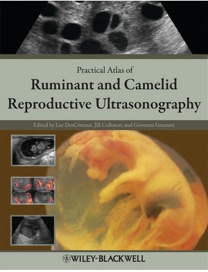
[P0000CWY] Practical Atlas of Ruminant and Camelid Reproductive Ultrasonography 
() 해외배송 가능
event
상품상세정보
ISBN: 9780813815510
Hardcover
244 pages
January 2010, Wiley-Blackwell
Description
Practical Atlas of Ruminant and Camelid Reproductive Ultrasonography is a practical, fully referenced, image-based guide to the essential concepts of reproductive ultrasound in domesticated ruminants and camelids. Providing information to enable practitioners to incorporate ultrasound service into their practices, the book also includes more specialized information for advanced techniques such as fetal sexing, embryo transfer, color Doppler, and others. Practical Atlas of Ruminant and Camelid Reproductive Ultrasonography is a must-have reference for ruminant and camelid practitioners, instructors, and students.
Table of Contents
Preface.
Acknowledgments.
Introduction.
Chapter 1 Principles and recommendations, essential concepts, and common artifacts in ultrasound imaging.
Description and practical recommendations in the choice of ultrasound equipment with a view to image quality.
General principles and essential concepts to improve image quality.
Common artifacts.
Chapter 2 Scanning techniques and common errors in bovine practice.
Description of scanning technique.
Manipulation of the probe.
Common errors.
Chapter 3 Anatomy of the reproductive tract of the cow.
Genital tract.
Descriptive terminology of the ovary and ovarian structures.
Chapter 4 Bovine ovary.
Endocrinology and ovarian structures in pubertal cows.
Ovarian anomalies and differential diagnosis.
Use of color Doppler to monitor blood flow.
Ultrasound use in reproduction synchronization protocols for dairy cattle: Two perspectives.
Chapter 5 Bovine uterus.
Ultrasound of the uterus during the estrous cycle and normal postpartum period.
Color Doppler sonography of the uterine blood flow.
Ultrasound of the postpartum abnormal uterus and vagina.
Chapter 6 Bovine pregnancy.
Morphologic embryonic and fetal development up to day 55.
Ultrasound landmarks of standard early pregnancy diagnosis.
Early embryonic and fetal death.
Twins.
Chapter 7 Bovine fetal development after 55 days, fetal sexing, anomalies, and well-being.
Fetal development after 55 days.
Ultrasound fetal sexing.
Fetal anomalies.
Fetal well-being during late pregnancy (normal gestation, compromised pregnancy, and clone).
Chapter 8 Bovine embryo transfer, in vitro fertilization, special procedures, and cloning.
Embryo donors.
Oocyte collection for in vitro fertilization.
Recipients.
Management of clone recipients.
Chapter 9 Bull anatomy and ultrasonography of the reproductive tract.
Ultrasound equipment and techniques.
Anatomy of the reproductive system.
Anomalies and ultrasonographic imaging of external and internal reproductive organs.
Chapter 10 Buffalo and zebu cattle.
Equipment and scanning techniques.
Major differences between bovine and bubaline species.
Pathology.
Congenital and hereditary defects.
Ultrasound services in buffalo and zebu.
Chapter 11 Sheep and goats.
Usefulness of ultrasonography in small ruminants.
Equipment and scanning techniques.
Ultrasound imaging of the female reproductive tract.
Endocrine and ovarian processes that comprise the normal estrous cycle and pregnancy.
Fetal count, age, and sex.
Pathological conditions in the female.
Ultrasonographic evaluation of the male genital system.
Common abnormalities of the testis.
Common abnormalities of the accessory glands.
Chapter 12 Camelids.
Usefulness of ultrasonography in camelids.
Equipment and scanning techniques.
Ultrasonographic anatomy.
Ovarian function and endocrinology in South American camelids.
Pregnancy diagnosis and evaluation of fetal growth.
Uterine and ovarian abnormalities.
Index.
Author Information
Editor-in-chief:
Luc DesCôteaux, DMV, MSc, Dipl. ABVP (Dairy), is a Full Professor and Service Chief of the Ambulatory Clinic at the Université de Montréal, Québec, Canada.
Associate Editors:
Giovanni Gnemmi, DVM, PhD, Dipl. ECBHM, is a bovine practitioner and ultrasound instructor with BovineVet Italy.
Jill Colloton, DVM, is a bovine practitioner and ultrasound instructor with Bovine Services, LLC, USA.
배송 정보
- 배송 방법 : 택배
- 배송 지역 : 전국지역
- 배송 비용 : 3,000원
- 배송 기간 : 2일 ~ 5일
- 배송 안내 : - 산간벽지나 도서지방은 별도의 추가금액을 지불하셔야 하는 경우가 있습니다.
고객님께서 주문하신 상품은 입금 확인후 배송해 드립니다. 다만, 상품종류에 따라서 상품의 배송이 다소 지연될 수 있습니다.
고객님께서 주문하신 도서라도 도매상 및 출판사 사정에 따라 품절/절판 등의 사유로 취소될 수 있습니다. 입금확인이 되면 익일발송(주말,공휴일제외)을 원칙으로 합니다.
교환 및 반품 정보
교환 및 반품이 가능한 경우
- 상품을 공급 받으신 날로부터 7일이내 내용이 파본일 경우
단, 비닐포장을 개봉하였거나 내용물이 훼손되어 상품가치가 상실된 경우에는 교환/반품이 불가능합니다.
- 공급받으신 상품 및 용역의 내용이 표시.광고 내용과
다르거나 다르게 이행된 경우에는 공급받은 날로부터 3월이내, 그사실을 알게 된 날로부터 30일이내
교환 및 반품이 불가능한 경우
- 고객님의 책임 있는 사유로 상품등이 멸실 또는 훼손된 경우. 단, 상품의 내용을 확인하기 위하여
포장 등을 훼손한 경우는 제외
- 포장을 개봉하였거나 포장이 훼손되어 상품가치가 상실된 경우
- 고객님의 사용 또는 일부 소비에 의하여 상품의 가치가 현저히 감소한 경우
- 시간의 경과에 의하여 재판매가 곤란할 정도로 상품등의 가치가 현저히 감소한 경우
- 복제가 가능한 상품등의 포장을 훼손한 경우
(자세한 내용은 고객만족센터 1:1 E-MAIL상담을 이용해 주시기 바랍니다.)
※ 고객님의 마음이 바뀌어 교환, 반품을 하실 경우 상품반송 비용은 고객님께서 부담하셔야 합니다.


