


- tel010-4724-3240
- fax02-447-3074
- time9시-18시

현재 위치


[P0000CXH] Radiography of the Dog and Cat: Guide to Making and Interpreting Radiographs 2013 

() 해외배송 가능
| 판매가 | |
|---|---|
| 소비자가 | 134,000원 |
| 적립금 |
|
| 무이자할부 | |
| 제조사 | |
| 원산지 | |
| 상품코드 | P0000CXH |
| 수량 |
|
| 국내/해외배송 | |
| SNS 상품홍보 | |
| QR코드 |
|
| QR코드 보내기 |
   
|


event
상품상세정보
소동물 방사선촬영 판독의 지침서입니다.
임상을 오래하신 원장님들은 기억하시겠지만 Jerry M. Owens의 Radiographic interpretation for the small animal clinician, 2/e(1998)과 비슷한 컨셉의 판독 지침서입니다.
일반 방사선학 서적과 달리 실제 방사선 사진은 많이 수록되지 않았읍니다.
대신 일러스트가 주로 수록되어 있으며 수록된 방사선 사진들은 이해를 돕기 위하여 강조처리가 되어 있읍니다.
한 가지 아쉬운 점은 내용의 구성이 로마숫자로 되어 있다는 점입니다.
국내 독자들에게 로마숫자로 일련번호가 매겨져 있으면 읽으면서 조금 걸리적 거리는 느낌이 있읍니다.
그간 발간되었던 방사선 사진 판독에 대한 서적들과 비교했을 때 수록된 내용의 양과 질 면에서 상당한 우위를 차지할 수 있을 것 같습니다.
또 한 가지 아쉬운 점은 최근 해외 대형 출판사들이 비닐포장을 하지 않는 경우가 잦아지고 있읍니다.
그 이유는 환경보호라고 하지만 그보다는 비용절감의 측면이 더 강한 것 같습니다.
아무튼!
이 문제로 인해서 도서의 표지 등에 약간의 잔흠집이 나 있는 경우가 많습니다.
이 점 양해 부탁드립니다.
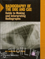
Radiography of the Dog and Cat: Guide to Making and Interpreting Radiographs
Radiography of the Dog and Cat: Guide to Making and Interpreting Radiographs offers a comprehensive guide to producing high-quality radiographs and evaluating radiographic findings. Equally useful as a quick reference or for more in-depth information on specific diseases and disorders, the book is logically organized into sections describing how to make high-quality radiographs, normal radiographic anatomy, and interpretation of radiographic abnormalities. It is packed with checklists for systematic evaluation, numerous figures and line drawings, and exhaustive lists of differential diagnoses, resulting in an especially practical guide for the radiographic procedures performed in everyday practice.
Written in a streamlined, easy-to-read style, the book offers a simple and fresh approach to radiography of the dog and cat, correlating physics, physiology, and pathology. Coverage includes patient positioning, contrast radiography, normal and abnormal radiographic findings, and differential diagnoses as they pertain to musculoskeletal, thoracic, and abdominal structures. Radiography of the Dog and Cat: Guide to Making and Interpreting Radiographs is a one-stop reference for improving the quality and diagnostic yield of radiographs in your clinical practice.
- Provides a practical guide to making and interpreting quality radiographs
- Includes complete evaluation checklists, extensive differential diagnoses, and numerous figures and line drawings
- Serves as both a fast access manual to key information and as a more detailed reference to specific diseases, disorders, and abnormal radiographic findings
- Discusses both analog and digital radiography systems
- Offers concrete guidance for improving the quality of radiographs and the accuracy of diagnosis
- Includes a companion website featuring review questions, a positioning guide, and figures and the glossary from the book at www.wiley.com/go/muhlbauerradiography
목차
About the Companion Website, ix
Acknowledgment, xi
Introduction, xiii
Chapter 1: Interpretation of Radiographs, 3
Musculoskeleton, 4
Thorax, 12
Abdomen, 27
Chapter 2: Making Quality Radiographs, 43
Production and use of diagnostic x-rays, 44
Positioning guide, 59
Interpreting radiographs, 82
Scope of diagnostic imaging, 85
Chapter 3: Contrast Radiography, 87
Indications for contrast radiography, 88
Types of contrast media, 88
Adverse effects associated with contrast media, 89
Contrast radiography procedures, 91
Chapter 4: Musculoskeleton, 123
Normal radiographic anatomy, 125
General, 125
Bone development, 128
Sesamoid bones, 130
Variable ossifi cation centers, 132
Joints, 133
Skull, 146
Diseases and disorders, 152
Bone production, 152
Bone loss, 156
Benign conditions, 157
Fractures, 161
Hypertrophic osteopathy (HO), 171
Osteomyelitis, 171
Osseous neoplasia, 174
Soft tissues, 177
Appendicular skeleton, 180
Axial skeleton, 232
Differential diagnoses, 264
General bone, 264
Joints, 267
Spine, 268
Skull, 271
Pharynx and larynx abnormalities, 273
Chapter 5: Thorax, 275
Normal radiographic anatomy, 277
Thoracic wall, 277
Diaphragm, 279
Pleura and pleural space, 279
Mediastinum, 280
Esophagus, 282
Heart and major vessels, 284
Trachea, 290
Bronchi, 290
Lungs, 291
Diseases and disorders, 294
Thoracic wall, 294
Diaphragm, 297
Pleura and pleural space, 299
Mediastinum, 304
Esophagus, 306
Heart and major vessels, 312
Trachea and bronchi, 335
Lungs, 341
Differential diagnoses, 356
Thoracic wall, 356
Diaphragm, 357
Pleura and pleural space, 358
Mediastinum, 360
Esophagus, 362
Cardiac silhouette, 364
Major vessels, 368
Trachea, 369
Lungs, 371
Chapter 6: Abdomen, 377
Normal radiographic anatomy, 379
Peritoneal and retroperitoneal spaces, 379
Liver, 380
Gall bladder/biliary system, 381
Spleen, 381
Pancreas, 383
Gastrointestinal tract, 383
Urinary tract, 388
Adrenal glands, 390
Male genital system, 391
Female genital system, 393
Diseases and disorders, 393
Peritoneal hernia, 393
Liver, 394
Gall bladder/biliary system, 396
Spleen, 397
Pancreas, 398
Gastrointestinal tract, 399
Urinary tract, 415
Adrenal glands, 427
Male genital system, 429
Female genital system, 433
Differential diagnoses, 436
Abdominal cavity, 436
Abdominal wall, 440
Gastrointestinal tract, 441
Liver, 447
Spleen, 448
Pancreas, 449
Urinary tract, 450
Male genital system 455
Female genital system 455
Glossary, 457
Bibliography, 473
Index, 475
저자 안내
M.C. Muhlbauer, DVM, MS, DACVR, is President and CEO of Veterinary Imaging Specialists, PC.
S.K. Kneller, DVM, MS, DACVR, is Associate Professor Emeritus of Veterinary Clinical Medicine at the College of Veterinary Medicine, University of Illinois at Urbana-Champaign.
배송 정보
- 배송 방법 : 택배
- 배송 지역 : 전국지역
- 배송 비용 : 3,000원
- 배송 기간 : 2일 ~ 5일
- 배송 안내 : - 산간벽지나 도서지방은 별도의 추가금액을 지불하셔야 하는 경우가 있습니다.
고객님께서 주문하신 상품은 입금 확인후 배송해 드립니다. 다만, 상품종류에 따라서 상품의 배송이 다소 지연될 수 있습니다.
고객님께서 주문하신 도서라도 도매상 및 출판사 사정에 따라 품절/절판 등의 사유로 취소될 수 있습니다. 입금확인이 되면 익일발송(주말,공휴일제외)을 원칙으로 합니다.
교환 및 반품 정보
교환 및 반품이 가능한 경우
- 상품을 공급 받으신 날로부터 7일이내 내용이 파본일 경우
단, 비닐포장을 개봉하였거나 내용물이 훼손되어 상품가치가 상실된 경우에는 교환/반품이 불가능합니다.
- 공급받으신 상품 및 용역의 내용이 표시.광고 내용과
다르거나 다르게 이행된 경우에는 공급받은 날로부터 3월이내, 그사실을 알게 된 날로부터 30일이내
교환 및 반품이 불가능한 경우
- 고객님의 책임 있는 사유로 상품등이 멸실 또는 훼손된 경우. 단, 상품의 내용을 확인하기 위하여
포장 등을 훼손한 경우는 제외
- 포장을 개봉하였거나 포장이 훼손되어 상품가치가 상실된 경우
- 고객님의 사용 또는 일부 소비에 의하여 상품의 가치가 현저히 감소한 경우
- 시간의 경과에 의하여 재판매가 곤란할 정도로 상품등의 가치가 현저히 감소한 경우
- 복제가 가능한 상품등의 포장을 훼손한 경우
(자세한 내용은 고객만족센터 1:1 E-MAIL상담을 이용해 주시기 바랍니다.)
※ 고객님의 마음이 바뀌어 교환, 반품을 하실 경우 상품반송 비용은 고객님께서 부담하셔야 합니다.
서비스 문의

상품 Q&A
상품에 대해 궁금한 점을 해결해 드립니다.
게시물이 없습니다


법인명(상호): 농경애니텍 143853 대표자(성명): 양경모 사업자 등록번호 안내: [206-92-98752] 통신판매업 신고 광진제1944
전화: 010-4724-3240 팩스: 02-447-3074 주소: 서울 광진구 자양동 226-24 선진빌딩 3층
개인정보관리책임자: 양경모(okck@vbooks.co.kr)
Contact okck@vbooks.co.kr for more information.
Copyright © 2010 농경애니텍 All rights reserved.